SCM

Presentation
The microscopy facility of the Campus Saint Germain des Prés (CSGP) offers access to photonic microscopy techniques (widefield, confocal, Airyscan, slide scanner) along with tools for image analysis and storage solutions. The facility is run by an operations manager and a scientific manager. The main decisions are made with the committee, representing each CSGP research unit and which is consulted approximately once a month. Located on the 4th floor of the UFR des Sciences Fondamentales et Biomédicales, the facility is open to the researchers, engineers, technicians and students of the building and to external teams/private companies. The ambition of the facility is to expand according to the users’ needs.
News
Team


Equipements
3i AxL Cleared Tissue LightSheet
– Room H422
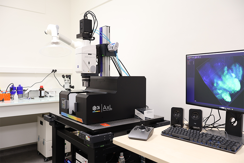
- Dual-sided light sheet illumination (thickness 2µm to 30µm)
- Motorized Zoom 0.7x-11.2x
- Compatible with a range of refractive indices (1.33 – 1.58)
- Chamber & Specimen Holders (Max Sample Dimensions : 25 x 25 x 20mm)
- Temperature Controlled Media
- Camera 1 : Hamamatsu ORCA-Fusion BT (2304 x 2304, 6.5µm)
- Software : SlideBook
| Lasers | Objectives | Emission Filterwheel |
| 405 nm 488 nm 561 nm 637 nm |
Illumination objectives: 2x 3i Custom Designed 0.14 NA Detection objectives: PlanNeoFluar Z 1.0x/0.25NA, WD : 56 mm PlanNeoFluar Z 1.5x/0.37NA, WD : 30 mm |
DAPI (445/45) FITC (525/30) TRITC (617/73) Cy5 (692/40) |
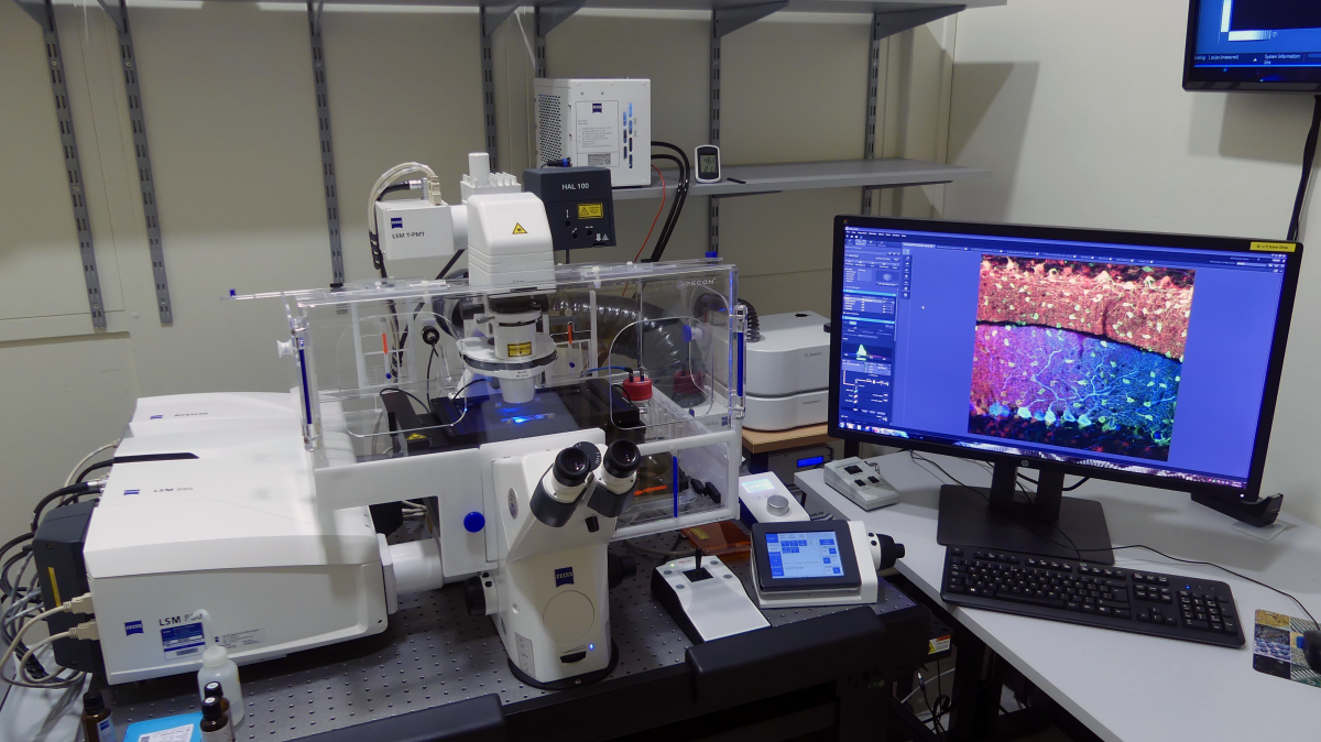
ZEISS LSM880 Fast Airyscan
– Room H423B
- High-resolution confocal
- 3D acquisition, time lapse, tiles, multi-positions, FRAP, spectral deconvolution
- Fixed/living samples
- Temperature and CO2 control
- Definite focus 2
- Software : ZEN Black 2.3
| Lasers | Objectives | Detection |
| 405 nm 445 nm 488 nm 514 nm 561 nm 594 nm 633 nm |
10x/0.45 Dry WD : 2 mm 20x/0.8 Dry WD : 0.55 mm 40x/1.2 Water Autocorr. WD : 0.28 mm 63x/1.4 Oil DIC WD : 0.19 mm |
2 PMTs 1 spectral PMT GaAsP 1 Airyscan detector GaAsP 1 Trans-PMT |
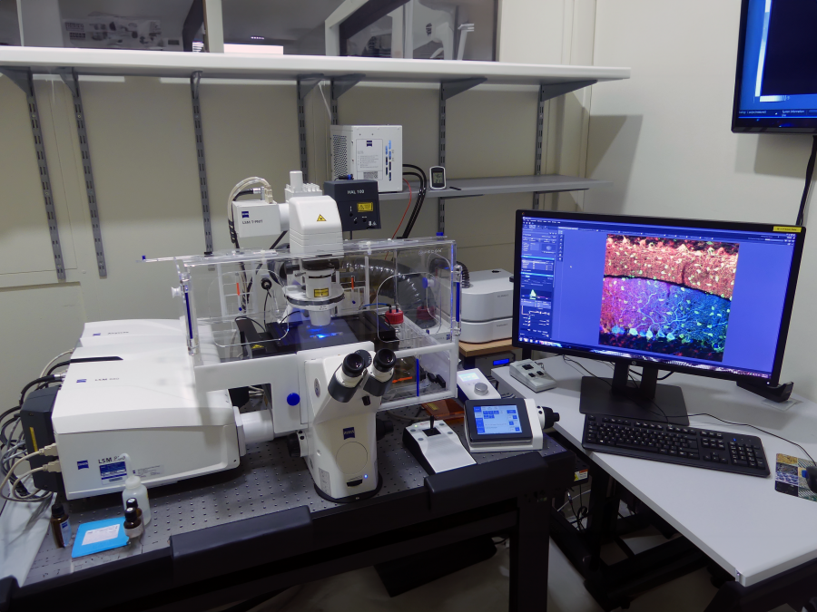
ZEISS LSM710 Confocal
– Room H423A
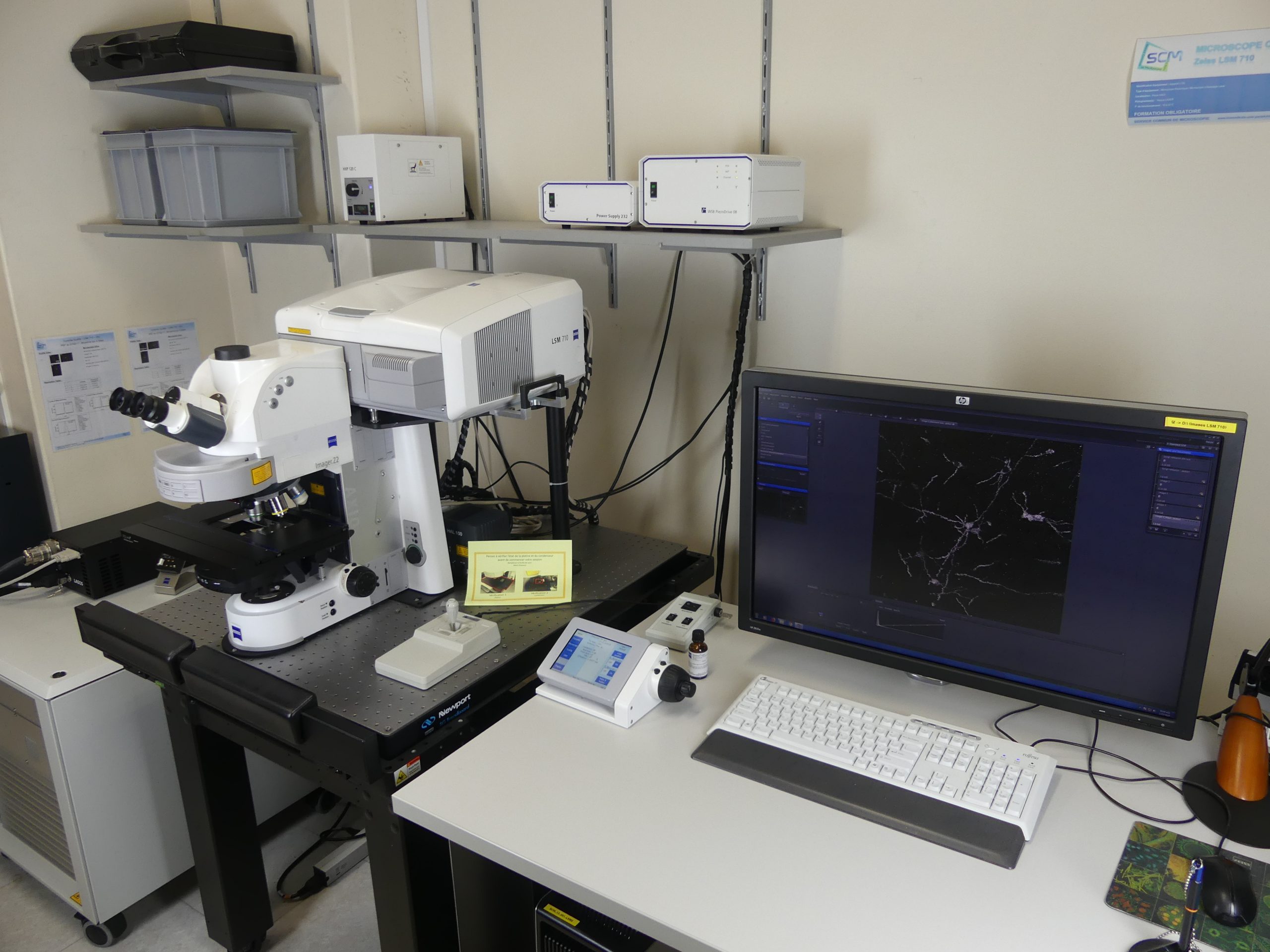
- 3D acquisition, time lapse, tiles, multi-positions, FRAP, spectral deconvolution
- Fixed/living samples
- Software : ZEN Black 2.1
| Lasers | Objectives | Detection |
| 405 nm 458 nm 488 nm 514 nm 561 nm 633 nm |
10x/0.45 Dry WD : 2 mm 20x/0.8 Dry WD : 0.55 mm 40x/1 Water DIC WD : 2.5 mm 63x/1.4 Oil WD : 0.19 mm 63x/1.2 Water WD : 0.28 mm |
2 PMTs 1 spectral PMT 1 Trans-PMT |
ZEISS LSM510 Confocal
– Room H423A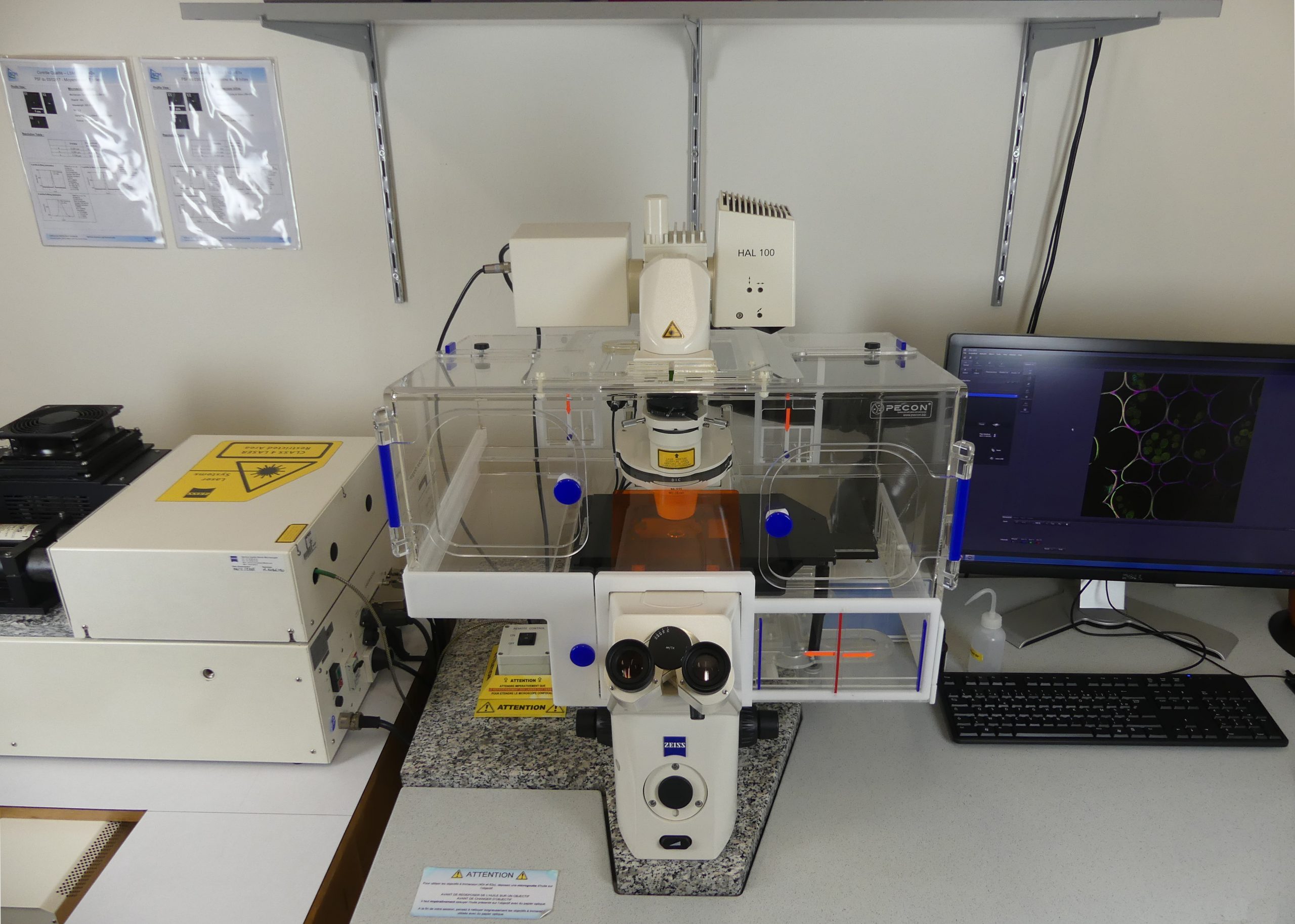
- 3D acquisition, time lapse
- Fixed samples
- Software : ZEN 2009
| Lasers | Objectives | Detection |
| 458 nm 477 nm 488 nm 514 nm 543 nm 633 nm |
2.5x/0.12 Dry WD : 6.3 mm 10x/0.3 Dry WD : 5.2 mm 20x/0.5 Dry WD : 2 mm 40x/1.3 Oil DIC WD : 0.2 mm 63x/1.4 Oil DIC WD : 0.19 mm |
2 PMTs 1 Trans-PMT |
ZEISS AxioScan.Z1 Slide scanner
– Room H423A
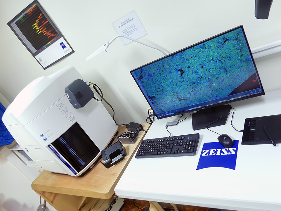
- 100 slides magazine, 3D acquisitions
- Fixed samples
- Brightfield, Fluorescence, Polarization
- Lightsource : Colibri 7 – LED System
- Camera 1 : Hamamatsu ORCA-Flash 4.0 (2048 x 2048, 6.5µm)
- Camera 2 : Hitachi HV F202 (1600 x 1200, 4.4µm )
- Graphic tab
- Software : ZEN 3.1
| LEDs | Objectives | Emission Filterwheel |
| 385 nm 430 nm 475 nm 511 nm 555 nm 590 nm 630 nm |
10x/0.45 Dry 20x/0.80 Dry 40x/0.95 Dry |
DAPI, Alexa Fluor 488 / 555 / 647 QBP : 425/30 + 514/30 + 592/25 + 709/100 CFP, YFP, mCherry TBP : 467/24 + 555/25 + 687/145 DAPI, FITC, mCherry TBP : 425/30 + 524/50 + 688/145 |
Nikon Eclipse TE-2000E Widefield microscope
– Room H438A

- 3D acquisition, time lapse, tiles, mutli-positions
- Brightfield/Fluorescence
- Fixed/living samples
- Temperature control
- Fluorescent lamp metal halide Nikon Intensilight 130W
- Camera 1 : Photometrics CoolSnap HQ2 (1392 x 1040, 6.45 μm)
- Camera 2 : QImaging Color Retiga 2000R (1600 x 1200, 7.4 μm)
- Software : NIS-Elements AR 3
| Objectives | Fluorescence cubes filters |
| 4x/0.13 Dry WD : 17.2 mm 10x/0.30 Dry WD : 16 mm 20x/0.75 Dry DIC WD : 1 mm 40x/1.30 Oil DIC WD : 0.2 mm 100x/1.45 Oil WD : 0.13 mm |
Dapi (377/50 D409 447/60) GFP (480/30 D505 535/40) TRITC (540/25 D565 605/55) Cy5 (628/40 D660 692/40) CFP (436/20 D455 480/10) YFP (500/20 D515 535/30) |
ZEISS Axioscope.A1 Widefield microscope
– Room H438A
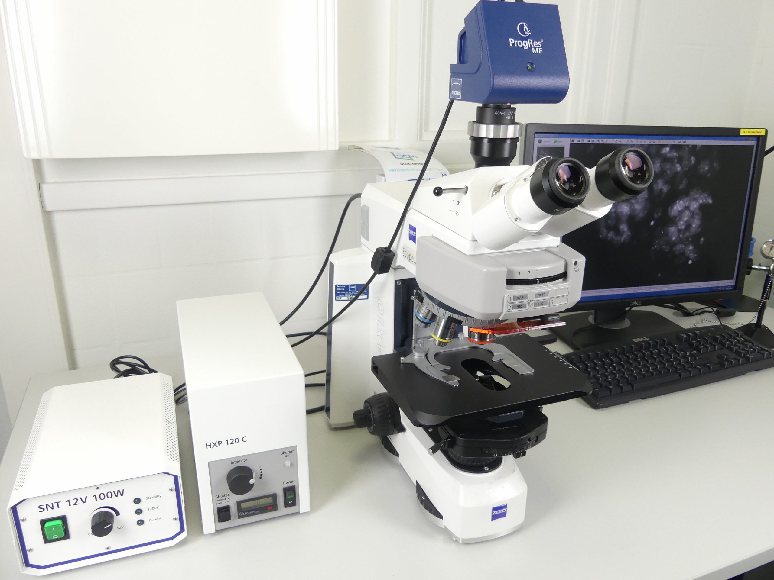
- Brightfield/Fluorescence
- Fixed samples
- Fluorescent lamp metal halide Zeiss HXP 120 C
- Camera 1 : Progres MF-USB Jenoptik (1360 x 1024, 6.45 μm)
- Software : ProgRes Capture Pro
| Objectives | Fluorescence cubes filters |
| 5x/0.16 Dry WD : 18.5 mm 10x/0.30 Dry WD : 5.2 mm 100x/1.4 Oil WD : 0.17 mm (sur demande 20x, 40x) |
Dapi (365 D395 445/50) FITC (475/40 D500 530/50) TRITC (545/25 D570 605/70) Cy5 (640/30 D660 690/50) |
Analysis Workstation 1
– Room H420B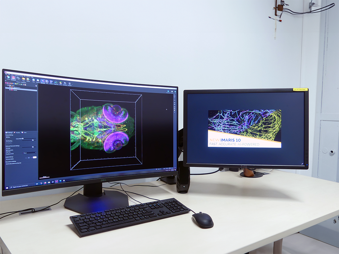
- AutoQuant X 3.1.3 Deconvolution software
- Ilastik 1.4.0
- QuPath v0.7.0
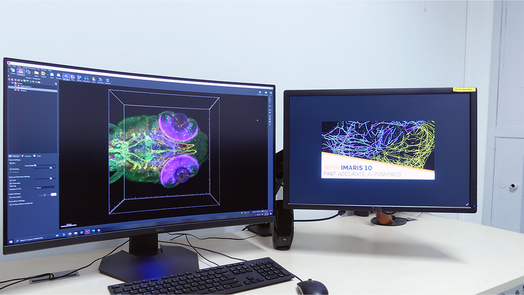
Analysis Workstation 2
– Room H420B
- Imaris 10 (Cell, Imaris XT, Coloc, Filament Tracer, Vantage, Batch)
- ZEN Blue lite
- ZEN Black lite (with Airscan processing module)
- NIS Elements AR Off-line
- ImageJ/Fiji
Laboratory fume cupboard – Sodatec
– Room H420A
- 3 micropipets Gilson (P20, P200, P2000) + pipet tips
- Dirt containers (sharp objetcts/paraformaldehyde)
- Precision Balance
- Digital Refractometer (DR301-95)
STORAGE SOLUTIONS AND POLICY
The images acquired on our systems are first stored on the acquisition computer. After each session on the systems, the user can transfer the files from the computer to our server (100 To). The images are stored on our computers for 6 months and for 3 years on our servers. After the delay, they are automatically deleted.
PRESTATIONS
The microscopy facility provides trainings on all the equipments mentioned above, under request. The autonomous users can ask to be assisted by a member of the facility if needed. We also provide our expertise and advices to guide our users in their experiments regarding sample preparation, if possible, imaging and image analysis.
PRICES
Prices 2026 (without engineer assistance)
| Equipements | Tarifs d’utilisation interne académique CSG (HT) | Tarifs d’utilisation externe académique Paris Cité (HT) | Tarifs d’utilisation externe académique (HT) | Tarifs d’utilisation privée (HT) |
| Sorbonne (Hotte Laboratoire) | Gratuit | Gratuit | Gratuit | Sur devis |
| Microscope Zeiss Primo Star | Gratuit | Gratuit | Gratuit | |
| Slide Scanner Zeiss AxioScan.Z1 | 7 € / Heure Scans longs nuit (17h-9h) : 40€ |
8 € / Heure Scans longs nuit (17h-9h) : 55€ |
11 € / Heure Scans longs nuit (17h-9h) : 80€ |
|
| Microscope WF 1 – NIKON Eclipse-TE2000E | 9 € / Heure | 9 € / Heure | 12 € / Heure | |
| Microscope WF 2 – ZEISS Axioscope A1 | Gratuit | Gratuit | Gratuit | |
| LightSheet – 3i Axl Cleared Tissue Lightsheet | Scans journée (4h), nuit (17h-9h) : 40€ | Scans journée (4h), nuit (17h-9h) : 45€ | Scans journée (4h), nuit (17h-9h) : 50€ | |
| Microscope Confocal 1 – ZEISS LSM 510 | 18 € / Heure | 20 € / Heure | 22 € / Heure | |
| Microscope Confocal 2 – ZEISS LSM 710 | 23 € / Heure | 24 € / Heure | 27 € / Heure | |
| Microscope Confocal 3 – ZEISS LSM 880 | 26 € / Heure | 32 € / Heure | 41 € / Heure | |
| Analysis WS 1 – Déconvolution | Gratuit | Gratuit | 5 € / Heure | |
| Analysis WS 2 – Reconstruction 3D | Gratuit | Gratuit | 7 € / Heure |
Contact / Access / Documention
Any question ? Please contact this address in order to have a better answser : scm@u-paris-sciences.fr / 01 76 53 10 77
Find the Microscopy Platform on the 4th floor of the Saints-Pères building and all the access details by following this link.
Charte SCM (PDF)
Remerciements UAR (PDF)
Partners

Publications
2024
- Triphenylamine Sensitized 8-Dimethylaminoquinoline: An Efficient Two-Photon Caging Group for Intracellular Delivery
Delphine Rigault, Philippe Nizard, Jonathan Daniel, Mireille Blanćhard-Desce, Eric Deprez, Patrick Tauc, Hamid Dhimane, Peter I. Dalko
https://doi.org/10.1002/chem.202401289 - The preferential injury of outer renal medulla after ischemia-reperfusion relies on high oxidative metabolism
https://doi.org/10.1101/2024.09.12.612245 - Satellite glial cells modulate proprioceptive neuron activity in dorsal root ganglia
Yasmine Rabah, Cendra Agulhon
https://doi.org/10.1101/2024.09.04.611156 - Understanding the Role of the SMN Complex Component GEMIN5 and Its Functional Relationship with Demethylase KDM6B in the Flunarizine-Mediated Neuroprotection of Motor Neuron Disease Spinal Muscular Atrophy.
https://doi.org/10.3390/ijms251810039
2023
- KCa1.1 channels contribute to optogenetically driven post-stimulation silencing in cerebellar molecular layer interneurons.
Kassa M, Bradley J, Jalil A, Llano I
J Gen Physiol. 2023 Jan 2;155(1):e202113004.doi: 10.1085/jgp.202113004. Epub 2022 Nov 3.
PMID: 36326690 - Virus-Free Method to Control and Enhance Extracellular Vesicle Cargo Loading and Delivery
Bui S, Dancourt J, Lavieu G
Bio Mater. 2023 Mar 20;6(3):1081-1091. doi: 10.1021/acsabm.2c00955. Epub 2023 Feb 13.
PMID: 36781171
2022
- A link between agrin signalling and Cav3.2 at the neuromuscular junction in spinal muscular atrophy
Perrine Delers, Delphine Sapaly, Badih Salman, Stephan De Waard, Michel De Waard & Suzie Lefebvre
Sci Rep . 2022 Nov 8;12(1):18960.doi: 10.1038/s41598-022-23703-x
PMID: 36347955 - Label-free, fast, 2-photon volume imaging of the structural organization of peripheral neurons and glia in the enteric ganglia
Doriane Hazart, 1,2,#,$ Brigitte Delhomme, 1 Martin Oheim 1,*,† and Clément Ricard 1
Front Neuroanat. 2023 Feb 9;16:1070062.doi: 10.3389/fnana.2022.1070062. eCollection 2022.
PMID: 36844894 - The cell polarity protein Vangl2 in the muscle shapes the neuromuscular synapse by binding to and regulating the tyrosine kinase MuSK
Myriam Boëx, Steve Cottin, Marius Halliez, Stéphanie Bauché, Céline Buon, Nathalie Sans, Mireille Montcouquiol, Jordi Molgó, Muriel Amar, Arnaud Ferry, Mégane Lemaitre, Andrée Rouche, Dominique Langui, Asha Baskaran, Bertrand Fontaine, Julien Messéant, Laure Strochlic
Sci Signal. 2022 May 17;15(734):eabg4982. Epub 2022 May 17.
PMID: 35580169
2021
- A simple, inexpensive and multi-scale 3-D fluorescent test sample for optical sectioning microscopies.
Olevsko I, Szederkenyi K, Corridon J, Au A, Delhomme B, Bastien T, Fernandes J, Yip C, Oheim M, Salomon A.
Microsc Res Tech. 2021 May 18. doi: 10.1002/jemt.23813. Online ahead of print.
PMID: 34008289
2020
- Complex context relationships between DNA methylation and accessibility, histone marks, and hTERT gene expression in acute promyelocytic leukemia cells: perspectives for all-trans retinoic acid in cancer therapy.
Garsuault D, Bouyer C, Nguyen E, Kandhari R, Prochazkova-Carlotti M, Chevret E, Forgez P, Ségal-Bendirdjian E.
Mol Oncol. 2020 Jun;14(6):1310-1326. doi: 10.1002/1878-0261.12681. Epub 2020 Apr 22.
PMID: 32239597 - The Small-Molecule Flunarizine in Spinal Muscular Atrophy Patient Fibroblasts Impacts on the Gemin Components of the SMN Complex and TDP43, an RNA-Binding Protein Relevant to Motor Neuron Diseases.
Sapaly D, Delers P, Coridon J, Salman B, Letourneur F, Dumont F, Lefebvre S.
Front Mol Biosci. 2020 Apr 17;7:55. doi: 10.3389/fmolb.2020.00055. eCollection 2020.
PMID: 32363199
2019
- Modulation of lung cancer cell plasticity and heterogeneity with the restoration of cisplatin sensitivity by neurotensin antibody.
Wu Z, Fournel L, Stadler N, Liu J, Boullier A, Hoyeau N, Fléjou JF, Duchatelle V, Djebrani-Oussedik N, Agopiantz M, Ségal-Bendirdjian E, Gompel A, Alifano M, Melander O, Trédaniel J, Forgez P.
Cancer Lett. 2019 Mar 1;444:147-161. doi: 10.1016/j.canlet.2018.12.007. Epub 2018 Dec 21.
PMID: 30583074 - Cisplatin increases PD-L1 expression and optimizes immune check-point blockade in non-small cell lung cancer.
Fournel L, Wu Z, Stadler N, Damotte D, Lococo F, Boulle G, Ségal-Bendirdjian E, Bobbio A, Icard P, Trédaniel J, Alifano M, Forgez P
Cancer Lett. 2019 Nov 1;464:5-14. doi: 10.1016/j.canlet.2019.08.005. Epub 2019 Aug 9.
PMID: 31404614 - A novel model of trauma-induced cerebellar injury and myelin loss in mouse organotypic cerebellar slice cultures using live imaging.
Llufriu-Dabén G, Meffre D, Massaad C, Jafarian-Tehrani M.
J Neurosci Methods. 2019 Jan 1;311:385-393. doi: 10.1016/j.jneumeth.2018.09.023. Epub 2018 Sep 22.
PMID: 30253198 - Mechanical Stretch of High Magnitude Provokes Axonal Injury, Elongation of Paranodal Junctions, and Signaling Alterations in Oligodendrocytes.
Chierto E, Simon A, Castoldi F, Meffre D, Cristinziano G, Sapone F, Carrete A, Borderie D, Etienne F, Rannou F, Morrison B 3rd, Massaad C, Jafarian-Tehrani M.
Mol Neurobiol. 2019 Jun;56(6):4231-4248. doi: 10.1007/s12035-018-1372-6. Epub 2018 Oct 8.
PMID: 30298339
Gallery