Retinal Organoid

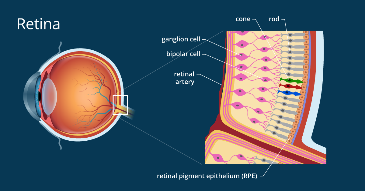
The retina is the ten-layered nervous tissue membrane of the eye. The outer layer, the retinal pigment epithelium (RPE), is a pigmented epithelial monolayer that ensures light absorption, nutrient transport, retinal storage for the visual cycle, phagocytosis and retinal attachment. The other 9 layers form the nerve retina (NR), which is responsible for the conversion of photons into a bioelectrical signal, its modulation and transmission, mainly to the visual cortex, by the optic nerve.
As part of the INOC competition, we have chosen to model the retina for its major medical issues in an aging society whose prevalence of retinal pathologies is gradually increasing. More than 170 million people are now affected by age-related macular degeneration (DMA) (Pennington & DeAngelis, 2016), a progressive deterioration of the area responsible for maximum visual acuity inducing a permanent spot in the center of the field visual, frequently occurring in 65 years old people.

In recent years, several models of retinal organoids with different levels of complexity have been developed, with a significant structural and functional variability within and between models. They reveal some difficulties in terms of yield, functionality (especially on photoreceptors) including the presence of non-retinal structures.
Our model aims to improve the regularity, reproducibility and functionality of the retinal Organoid. Thus, to create this Organoid, we will reproduce the steps of Embryonic development of the retina by differentiating murine embryonic stem cells and induced human pluripotent cells (IPS). They will be cultured in a microfluidic system, to allow miniaturization and automation of cell culture procedures, enabling us to fulfill our objective of optimised reproducibility.


